The Power of Microscopy: Applications in Science, Industry, and Beyond
What is Microscope?
Microscope is an instrument that helps to enlarge a small object so that it becomes seen under the naked eye. It is quite effective in helping us analyze the underrating structures of an object and we would not be able to see this with our naked eye. From the complexity of cellular kingdoms to the construction of crystals, the lens of microscope has undergone a tremendous distance in transforming the way people perceive the concepts existing around them.
Historical Context:
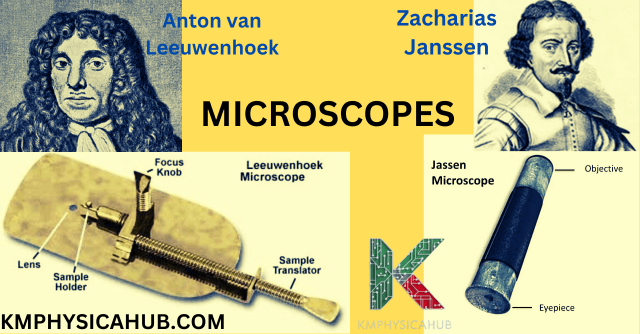
Microscope in terms of a device and an invention can be traced back several hundred of years. The first microscope was constructed in the last decade of the sixteenth century and the builder of the first compound microscope was a spectacle maker named Zacharias Janssen from the Netherlands. However, it was Anton van Leeuwenhoek, a Dutch scientist who revolutionized the microscopy in the 17th century. His well-calculated single lens optical microscopes with a magnification power of 200 times allowed him to make a first analysis of bacteria, blood corpuscles, and other small living creatures.
English scientist Robert Hooke elaborated this approach with his compound microscope, where he published observations of plants and insects in the “Micrographia”. These pioneers set the basis for today’s microscopy.
Types of Microscopes
Light Microscopes:
The light microscopes utilize visible light to illuminate the specimen and also to magnify it. They are broadly categorized into:
- Compound Microscopes:
These microscopes are those which have the capacity to use several lenses to make the image even larger. These accessories are widely employed in different educational institutions and research facilities.
- Stereo Microscopes:
Also referred to as dissecting microscopes, these microscopes offer depth vision pictures of the specimen, and so are suitable when dealing with large specimens such as insects or tissues on plants.
Electron Microscopes:
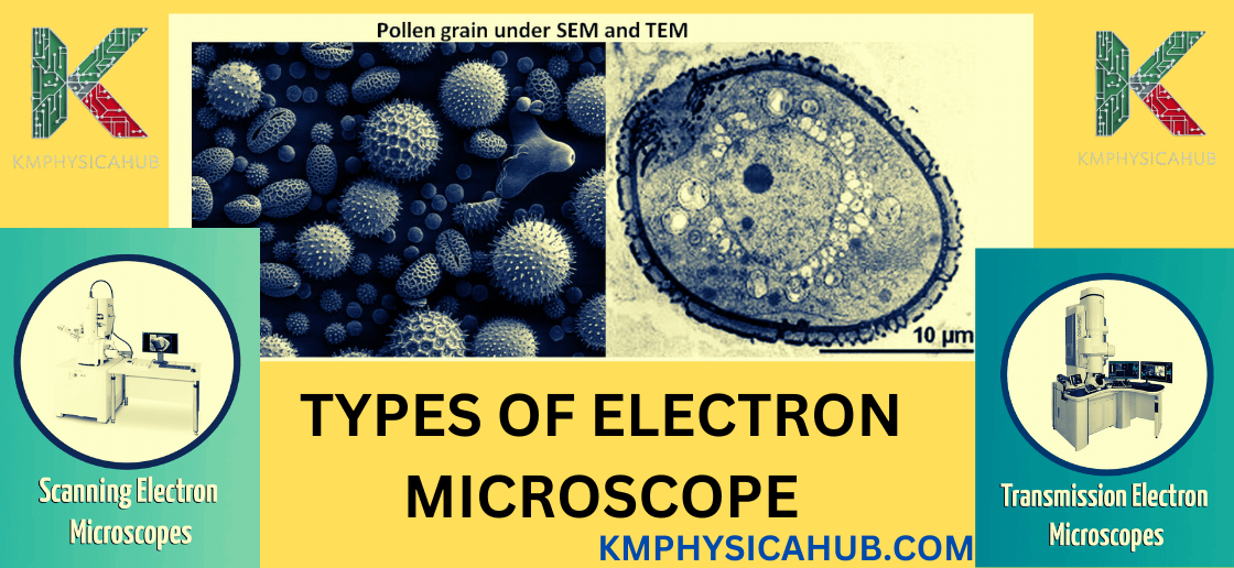
Unlike in the case of light microscopes, electron microscopes use electron beam to light up the specimens. They offer bigger resolution and resolving capability compared with the other light microscopes and are capable of showing us the internal structural aspect of a cell and the materials.
- Scanning Electron Microscopy (SEM):
SEM operates like a stereo microscope that zones in to some depth, obtaining the 3D image of the top layer after the surface was scanned with a thin beam of electrons.
- Transmission Electron Microscopy (TEM):
TEM is different in that it only uses a very thin slice of the sample to be illuminated by a beam of electrons! Hence, it gives an incredible clear look of the inside of the specimen through the electrons shot at the sample.
Other Microscopy Techniques:
Beyond light and electron microscopes, several specialized techniques have emerged to address specific research needs:
Confocal Microscopy: Under this technique, the sample is exposed to laser and the reflected lasers are used to produce images of specific layers within the specimen.
Fluorescence Microscopy: This technique involves the use of probes that have the ability to produce structural images of the specimen or detect some molecules when subjected to specific wavelengths of light.
Key Components of a Microscope
Objective Lens:
The objective lens is the one that focuses light on the specimen to provide the first level of enlargement. It’s positioned nearest to the specimen and is often available in several powers (e. g., 4x, 10x, 40x, 100x). It implies that the larger the value of magnification, the more details one gets to see on the sample.
Eyepiece:
These are the lenses through which one looks directly in order to see the enlarged image of the object. Additionally, the image that was formed by the objective lens is further enlarged by the eye lens. The overall power of the microscope depends on the conditional magnification of the objective lens and eyepiece.
Stage and Illumination:
A specimen is put on the stage which is a part of the structures of a microscope. It is usually fitted with clips to retain the specimen after insertion. The illumination source commonly a lamp is the unit that supplies the required light to the specimen. The type of illumination required also differs from one microscopy and the specimen for another.
Focusing Mechanisms:
The coarse and fine tuning knobs are utilized for moving the stages and specimen in order to optimally focus them. Coarse adjustment knob is used to get a rough focusing while the fine focus knob is used to increase the focus sensitive movements.
Applications in Biology
-
Cell Biology:
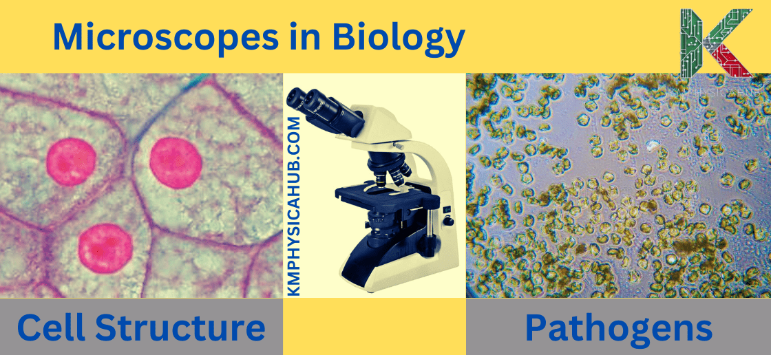
Microscopes enable identification of features of cells, which are the fundamental units of all living organisms, including their size, shape, functions, and components. As a result, we also come to know about the effects of light on plant’s growth through microscopy.
Observing Cell Structure: Compound microscopes and light microscopes do show the fundamental parts of cells such as the nucleus, cytoplasm and cell wall. Again, electron microscopes offer a even higher level of magnification enabling one to see cellular structures such as mitochondria, golgi apparatus and endoplasmic reticulum among others.
Studying Cell Processes: With the help of microscopes, details of dynamic processes occurring within cells such as mitosis and meiosis, synthesis and transport of protein can be clearly visualized.
Investigating Cellular Pathology: Through microscopes, various characteristics of cells related to disease can be seen, and knowledge can be gained about the disease and its treatment.
-
Microbiology:
Microscopes are essential tools in microscopic biology, for visualizing microorganisms, such as bacteria, viruses, fungi, and protozoa.
Identifying Pathogens: Microscopes help in differentiating and categorizing infections particles which aid in disease diagnosis and management.
Understanding Microbial Ecology: Microscopes observe and explain the distribution and relationships of microbes in environments like soil, water and the human body.
Developing Antimicrobial Agents: The use of microscopes, particularly in laboratories, is paramount when investigating the impact that antibiotics or any other antimicrobial substance might have on the growth and proliferation of microbial species.
-
Genetics:
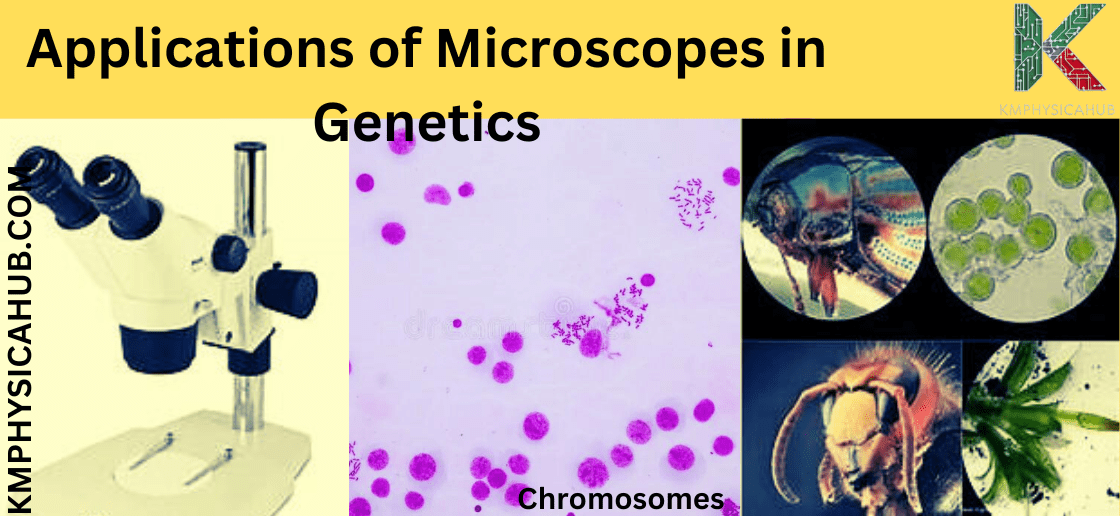
Microscopes are essential in genetics since they enable an examination of the DNA and its components, including the genes, chromosomes, and more.
Analyzing Chromosomes: Microscopes help to observe chromosomes, to investigate their properties, number and features of the chromosomal abnormalities that cause genetic disorders.
Investigating Gene Expression: Molecular fluorescent microscopy is a practical tool in observing expression of particular genes in cells, thus studying gene regulation and other cellular activities.
Developing Gene Editing Techniques: Microscopes are used in the process of creating genetic engineering technologies such as the CRISPR-Cas9 which can modify genes in living organisms.
-
Medicine:
Operating microscopes are crucial in medical practice as they are used in diagnosis, planning for treatment procedures and research.
Diagnosing Diseases: It is used for imaging tissue samples, blood smears, or any other biological material to diagnose diseases or diagnose an abnormally functioning cell.
Identifying Pathogens: Microscopy assists in specifying certain bacteria or virus that needs to be treated with certain antibiotic or antiviral.
Developing New Treatments: Microscopes help in understanding the action of new drugs and treatments on cells and tissues, which would help in the formation of more effective drugs.
Applications in Material Science
-
Nanotechnology:
Nanotechnology is a field of science that deals with studying, manipulating and using substances at the level of atoms and molecules. In order to achieve this, microscopes are essential tools since they allow us to see these particles.
Characterizing Nanomaterials: In order to characterize nano-sized materials, electron microscopes like TEM are utilized for analyzing their structure, morphology and characteristics.
Developing Nanotechnologies: It also helps in developing new technologies for various applications including electronics, energy harvesting systems and medical equipment by enabling researchers to investigate nanoscale devices.
Investigating Nanomaterial Interactions: The use of microscopes can also lead to understanding how nanomaterials work within living organisms that may eventually pave way to smart drug delivery systems or any other biomedical areas.
-
Material Characterization:
Microscopes are also used to study the structure, composition, and other characteristics related to the behavior and properties of materials.
Analyzing Material Structure: Optical microscopes are essential for understanding the atomic and molecular structures of materials, including their crystal structure, regularities, and inclusions.
Determining Material Composition: Microscopes can also identify the composition of materials, revealing which elements or chemical compounds they consist of.
Investigating Material Properties: Microscopes aid researchers in analyzing the mechanical, electrical, optical, and other properties of materials, leading to the development of new and improved high-performance materials.
-
Quality Control:
Microscopes are used in quality assurance and control, in determining the quality/standard of materials used during manufacturing.
Inspecting Material Defects: In applications like detecting flaws, contaminants or any other abnormalities in a material, microscopes prove quite useful to assess the quality levels of items.
Monitoring Material Processing: Microscopes are used in checking the processed material to see if the required properties have been met.
Ensuring Product Quality: Finished products are also viewed under microscopes to confirm if they are of the right quality and do not have any any flaws.
Applications in Forensic Science
Today, microscopes play an important role in forensic activities and help in investigating the criminal cases.
Analyzing Fibers and Hairs: Microscopes enable them to analyze fibers and hairs that are likely to be left behind at the scene of a crime to make conclusions of who could have committed the crime or where the material used in the commissioning of the crime could have been sourced from.
Examining Firearms Evidence: Bullet fragments and cartridge cases are also analyzed with microscopes to get details about the firearm, as well as possible connections to other cases.
Analyzing Trace Evidence: There are other forms of traces that can be analyzed using microscopes, such as paint, glass, and soil particles that may prove useful in connecting criminals to a crime scene.
Applications in Archaeology
In archaeology, microscopes are useful in analyzing a part of the relics or fossils for understanding the lost cultures and history of the evolution of life forms.
Examining Ancient Artifacts: Microscopes help archaeologists in determining the origin of the artifacts, the techniques in their making and other technicalities of the artifacts which can act as a window to the past and the cultures of people who made these artifacts.
Analyzing Fossils: Microscopes that are employed in analyzing the morphology and the internal structure of fossils are very helpful in painting the picture of evolution and history of life on Earth.
Revealing Hidden Details: Using microscopes on artifacts and fossils can also help to observe surface writings, wear and damages, as well as the underlying structures essential for study and analysis.
Applications in Industry
Microscopes in many industries are effectively used for controlling the quality of products, monitoring production and creation of new technologies.
Quality Control in Manufacturing: Products require inspection for defects and dimensional control and microscopes offer a way to monitor the quality of the materials input to the processes.
Semiconductor Production: Microscopes are very critical in semiconductor industry mostly for the purposes of inspecting and maneuvering different small parts and circuits.
Research and Development: An excellent example of general business application of microscopes is in the research and development industries where microscopes are used to study the properties of new materials and for developing new products and enhancing existing manufacturing procedures.
Advancements in Microscopy
Conventionally, the diffraction limit of light has been a constraint in microscopy and has in effect stunted the advancements of light microscopes for quite a long period of time. However, the above view has been debunked with the onset of recent technologies in a field referred to as super-resolution microscopy which makes it possible to image objects that are smaller than the wavelength of light.
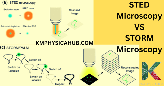
- Stimulated Emission Depletion (STED) Microscopy:
This technique relies on the utilization of the focused laser beam in order to selectively excite fluorescence molecules and so, reduce the area of the sample illuminated and thus, obtain a higher spatial resolution.
- Stochastic Optical Reconstruction Microscopy (STORM):
This is done using probes that can be ‘activated’ to produce either low or high resolution; from which a final high resolution image can be reconstructed using fragments of the unit probes.
- Photoactivated Localization Microscopy (PALM):
Similar to STORM, PALM uses fluorescent probes but in this case, it only activates and confines the fluorophore to the area of interest which requires high magnification.
Future Directions
The field of microscopy is constantly evolving, with ongoing research focused on developing even more powerful and versatile techniques:
Light Sheet Microscopy: It employs a sheet of light to illuminate the specimen and has low phototoxicity to enable fast three-dimensional imaging of live specimens.
Multiphoton Microscopy: This technique stems from the use of longer wavelengths of light to give better penetration into the sample and allow imaging through thick samples.
Correlative Microscopy: Applying multi-scale microscopy simply means that the specimen is examined using these microscopies at different resolutions as offered by the techniques.
Conclusion:
The use of microscopes in physics has played a key role in how we see the universe and appreciate the miracles of the ‘little world’. Microscopy has a history that dates back to the early single lens microscope, to the definition of ‘new’ techniques today, microscopy is always a fantastic tool for human beings in order to expand the view of the world beyond what we actually cannot see.
The opportunities remain numerous with further developments paving the way to more discoveries about the mechanisms of life and existence. Thus, microscopes will remain valuable resources in the ongoing quest to investigate deeper into the minute wonders of our earth and the rest of the expanding universe.
FAQS
Q1. What is the diffraction limit of light?
A: It’s the predicted boundary in light microscopes that cannot be broken due to the light’s wavelength. Nevertheless, super-resolution methods manage to overcome this obstacle.
Q2. What are the different types of microscopes?
A:
Light Microscopes: Use visible light to illuminate and magnify the specimen.
Electron Microscopes: Use an electron beam to visualize an image, which provides a higher level of detail than conventional light microscopes.
Q3. What is the difference between a compound microscope and a stereo microscope?
A: Compound microscopes: The main working principle of a compound microscope involves passing light through a series of lenses that refract the light and produce a larger image of an object. This technology enables the examination of minute details.
Stereo microscopes: Simultaneously the user also achieves a 3D effect because of the existence of two separate lenses which the noise is finally rectified onto as one. It appears to the user as if the object is three-dimensional and as if it is right in front of him/her.
Q4. How does magnification work in a microscope?
A: When we discuss a microscope and the purpose of magnification, it should be emphasized that the lens closest to the specimen bends light rays toward the internal lenses, thus magnifying the image of the object from a distance. Higher magnification means the larger the image.
Q5. What is resolution in microscopy?
A: Resolution is the shortest distance between two objects that keeps them from being seen as one. A microscopy with higher resolution will reveal the finest details.
Q6. What is a microscope?
A: Microscope, in science, an instrument used to produce larger images of objects that are too small to be seen with the naked eye. It is equipped with the lenses that can be used to refract and thereby, produce enlarged objective images.